

Revolutionizing Surgical Imaging: The Power of 3D-Printed Tumor Models
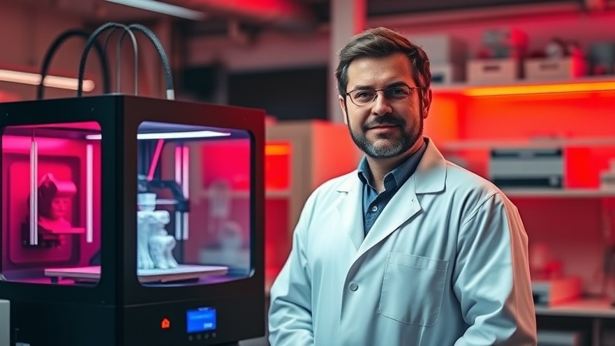
The field of surgical imaging has taken a groundbreaking turn with the innovative use of 3D-printed tumor models. Developed by Indrajit Srivastava and his team at Texas Tech University, these models offer a new lens through which health practitioners can refine surgical procedures, particularly tumor removals. By mimicking the optical and physical properties of actual human tumors, these phantoms pave the way for enhanced imaging techniques that promise to improve patient outcomes.
Understanding the Technology Behind Tumor Models
At the heart of this advancement lies the concept of 3D bioprinting, a technology that enables the construction of lifelike phantoms using materials like lipids, enzymes, and tumor cells combined with gelatin. By replicating the complexity of human tumors in size and composition, researchers can simulate real-world conditions during surgical imaging, leading to more reliable outcomes. This contrasts significantly with traditional practices requiring animal models, which may not accurately reflect the unique attributes of human physiology.
The Promise of Afterglow Imaging
One of the most exciting applications of these 3D-printed models is their role in afterglow imaging, a promising technique for tumor resection. According to Srivastava, afterglow imaging outperforms conventional fluorescence-guided approaches, allowing surgeons extended visibility into the tumor tissue for up to ten minutes post-exposure to light. This extended visibility is crucial, especially as it provides the surgeon with ample time to ensure accurate removal of the tumor without affecting surrounding healthy tissues.
The Challenge of Animal Models
As noted by Srivastava, traditional animal testing methods face substantial limitations. Mice, which are often utilized in these experiments, have skin that is significantly thinner than that of humans, leading to discrepancies in results that may not be applicable in the operating room. This raises the question: how can surgeons trust the efficacy of imaging agents tested on animals when human patients have complex layers that impact how these agents behave?
A Cost-Effective Alternative
Incorporating 3D-printed tumor models within surgical studies also presents significant economic advantages. Not only do these models reduce reliance on animal testing, but they also allow for extensive experimentation at a fraction of the cost. Health practitioners can achieve statistical significance in their data more efficiently, ultimately contributing to quicker advancements in surgical technologies.
Enhancing Surgical Precision with Real-World Applications
The potential for these tumor models extends beyond research laboratories. As they become integral to instructional simulations and real-time imaging, the broader implications suggest improvements in surgical precision. This could translate to shorter recovery times for patients, reduced hospital stays, and overall better health outcomes.
Future Insights: What Lies Ahead?
Looking forward, the next step for Srivastava and his team is to conduct comprehensive evaluations of these models in live settings, although they currently face hurdles, such as achieving widespread acceptance for afterglow imaging. However, the momentum of technology in healthcare continues to rise, and as more practitioners become aware of these breakthrough models, we may see rapid integration into standard medical protocols. By keeping an open dialogue about the latest advancements, concierge health practitioners can ensure they remain at the forefront of medical innovation.
The Takeaway for Health Practitioners
Concierge health practitioners play a vital role in connecting patients with cutting-edge treatments and technologies. Staying updated on innovations such as 3D-printed tumor models can help practitioners anticipate changes that may impact their services significantly. Knowledge is power—by understanding current advancements, practitioners can better advocate for their patients and incorporate the latest strategies into their care plans.
Embrace the future of surgical imaging. Stay informed about ongoing research and consider how these technologies could enhance your practice. The time for transformation is now—align your patient offerings with the latest innovations for the highest standard of care.


Write A Comment