

Revolutionizing Deep Tissue Imaging: The Power of Dual Wavefront Correction
In the quest for clearer insights into human biology, researchers have made striking advances in deep tissue imaging, which can significantly enhance patient diagnostics and treatment options. A groundbreaking study from the Technion – Israel Institute of Technology introduces dual wavefront correction, a technology that addresses past limitations in imaging depth and clarity. As wavefront shaping technology evolves, it offers promising solutions to common challenges faced by healthcare providers.
Moving Beyond Traditional Imaging Techniques
Historically, deep tissue imaging required the use of fluorescent points that had to be manually inserted into tissues. This invasive technique not only introduced potential risks but also impeded the ability to accurately visualize tissue without noise interference. These challenges made it clear that a more refined method was necessary.
Now, researchers have turned the spotlight on dual wavefront correction, enabling the direct imaging of neurons labeled with the fluorescent protein EGFP. This allows deeper visualization of biological structures, opening new doors for clinical applications.
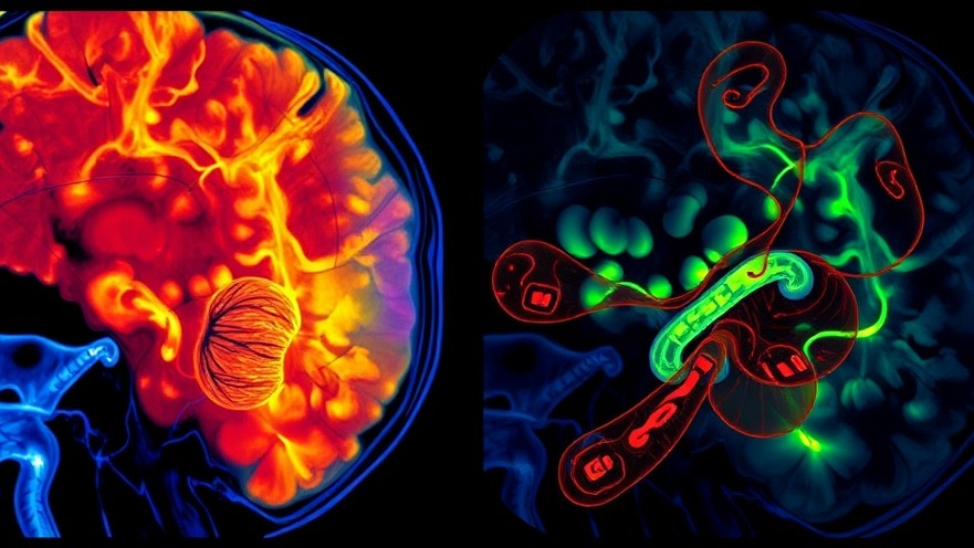
A Leap Forward: Understanding the Technology
The core of this innovation lies in simultaneously correcting the outgoing wavefront— the light sent into the tissue—and the incoming wavefront, which is the light returning from the tissue. By leveraging advanced mathematical models that optimize the signal-to-noise ratio, Dror Aizik and his team achieved unprecedented high-resolution images of neurons deep within the tissue.
What sets this technology apart is its capacity to overcome substantial distortions, essential for obtaining clearer visuals in complex biological environments. For healthcare practitioners, this could mean more accurate diagnoses, especially for conditions involving intricate neural networks.
The Implications for Healthcare: A New Era of Diagnostics
For concierge health practitioners, integrating this innovative imaging technique could revolutionize their diagnostic capabilities. The ability to visualize neurons and their axons from deeper layers of tissue will lead to higher precision in monitoring and diagnosing neurological conditions, ultimately enhancing patient outcomes.
Examining Wider Applications in Various Tissues
While the initial demonstrations focused on neuronal tissues, researchers believe that the benefits of dual wavefront correction extend to a variety of other tissues as well. This versatility opens the door to non-invasive imaging techniques across different medical specialties, from oncology to cardiology, showcasing the technology's potential to transform the entire healthcare landscape.
Future Trend: Non-Invasive Techniques Become Mainstream
The demand for non-invasive diagnostic methods has been steadily growing, and technologies like dual wavefront correction are pivotal in responding to this need. By minimizing invasiveness while maximizing visual clarity, healthcare providers can offer improved patient experiences and outcomes.
Making Informed Choices: What Practitioners Should Consider
Integrating new technologies into practice is no small feat. Health practitioners should thoroughly evaluate the reliability, costs, and staffing implications of adopting dual wavefront correction systems. As technology progresses, staying informed about these advancements ensures healthcare providers remain at the forefront of medical innovation.
Conclusion: Embracing Future Technology for Better Care
As wavefront shaping technology continues to evolve, it presents exciting possibilities for enhanced deep-tissue imaging capabilities. Practitioners should consider how to incorporate these advancements into their practices to provide unparalleled care for their patients. To stay ahead in this ever-changing landscape, explore options for integrating dual wavefront correction technology into your offerings.


Write A Comment