 Add Row
  Add
Add 

Revolutionizing Vascular Treatments with Bioprinting
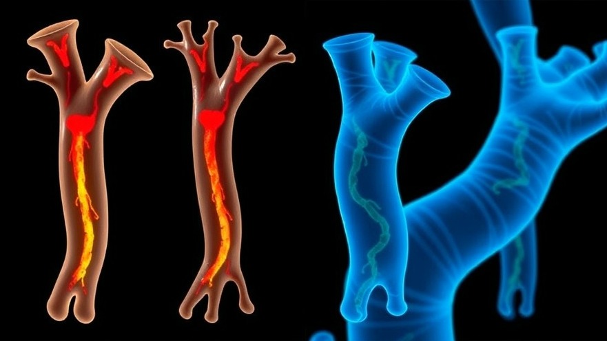
The recent breakthrough by researchers at Yale School of Medicine, who successfully implanted bioprinted synthetic aortas into rats, could signal a dramatic shift in how we approach vascular repairs and treatments for cardiovascular diseases. This innovative technology promises not only to enhance the efficacy of treatment methods but also to provide patients with custom-designed solutions that align intimately with their specific anatomical needs.
Understanding the Bioprinting Process
Bioprinting involves the use of 3D printing technology to create tissues or organs using living cells. In this study, scientists cultivated essential aortic cells, including smooth muscle cells and fibroblasts from rats, which were then printed to create a synthetic aorta. This innovative technique, contrasting sharply with traditional grafting methods, minimizes the time required to produce viable tissues, significantly accelerating the preparation phase that previously took weeks in bioreactors.
Potential Impact on Cardiovascular Treatments
Current methods for treating severe cardiovascular diseases often involve the use of autologous grafts made from the patient’s own tissue or synthetic materials, each of which carries inherent drawbacks. Autologous grafts, while effective, necessitate invasive surgeries and carry a risk of failure. Synthetic grafts, on the other hand, are typically only suitable for larger vessels and often face issues like leakage and bacterial infection. By replacing conventional methods with bioprinted vasculature, patients might experience not only more successful surgeries but also a healthier, more active recovery period.
What This Means for Health Practitioners
As concierge health practitioners, staying informed about groundbreaking developments like these is crucial to providing optimal patient care. The implications of bioprinting technology extend beyond surgical settings into patient education. Practitioners can educate patients about newer, less invasive options that might significantly improve their quality of life.
Future Directions for Research and Implementation
This proof-of-concept study showcases the potential of 3D bioprinting for broader applications in regenerative medicine. It opens avenues for further research into bioprinting not only the aorta but other critical arteries. Future studies will need to address longer-term outcomes and biocompatibility in larger animal models or eventual human trials to assess the robustness of bioprinted vascular structures. Regenerative medicine is at a pivotal intersection, and continued research could yield applications that redefine standards of care across cardiology.
Common Misconceptions About Bioprinting
Despite the promise bioprinting holds, several misconceptions linger regarding its feasibility and effectiveness. One common myth is that bioprinting can only be used for large organs. In reality, ongoing advancements suggest it can effectively produce smaller, more complex tissues, including blood vessels. Furthermore, people often underestimate the enormous precision and customization possible with this technology.
Concluding Thoughts and Call to Action
The successful implantation of bioprinted aortas is just the beginning. As we stand on the brink of significant advancements in cardiovascular solutions, now is the time for health practitioners to actively engage with the emerging technology trends in medicine. Explore ways to integrate bioprinting into your practice, and stay updated on future developments that may directly impact patient care. Educate your patients about the possibilities of bioprinted solutions and inspire them to embrace the future of personalized healthcare.


Write A Comment